2. ELECTRIC CURRENT FIELD
3. APPLICATIONS
![]() Effective Cells: all types of eukaryotic cells including all cell lines, primary cells, stem cells, blood and immune related cells, neurons, fetus cells and plant cells.
Effective Cells: all types of eukaryotic cells including all cell lines, primary cells, stem cells, blood and immune related cells, neurons, fetus cells and plant cells.
![]() Effective Biomolecules: DNA, RNA, siRNA, peptides and proteins
Effective Biomolecules: DNA, RNA, siRNA, peptides and proteins
![]() Applicable Experiments: transient expression, stable expression, gene knock-in/knock-out, siRNA transfection.
Applicable Experiments: transient expression, stable expression, gene knock-in/knock-out, siRNA transfection.
![]() Fields of Study: molecular and cellular biology, immunology, hematology, neuroscience, cancer research and drug discovery.
Fields of Study: molecular and cellular biology, immunology, hematology, neuroscience, cancer research and drug discovery.
4. Nucleus transfection is a wrong concept
Certain electroporation company has claimed that plasmids can be delivered into the cell nucleus directly. The “direct-to-nucleus” concept is wrong as revealed by at least three main reasonings.
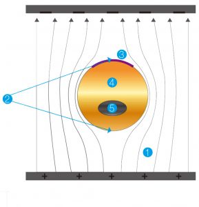
![]() (1) Electric current field distribution analysis: the cell membrane shoulders most of the electric potential applied on the cell and inside the cell, there is very little electric current or field distributed. The low internal potential is further distributed on the cell plasma and the cell nucleus, therefore the cell nucleus only gets a portion of the low electric field. Cell nucleus is smaller than a cell and require a higher potential to drive plasmids across, therefore it’s not possible for the internal electric field to be high enough for plasmid delivery directly into the nucleus. However, this is not an issue since plasmids can travel by themselves in the cell. Furthermore, this electric field distribution is very advantageous for the integrety of the genome during electroporation.
(1) Electric current field distribution analysis: the cell membrane shoulders most of the electric potential applied on the cell and inside the cell, there is very little electric current or field distributed. The low internal potential is further distributed on the cell plasma and the cell nucleus, therefore the cell nucleus only gets a portion of the low electric field. Cell nucleus is smaller than a cell and require a higher potential to drive plasmids across, therefore it’s not possible for the internal electric field to be high enough for plasmid delivery directly into the nucleus. However, this is not an issue since plasmids can travel by themselves in the cell. Furthermore, this electric field distribution is very advantageous for the integrety of the genome during electroporation.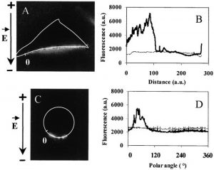
![]() (2) Visual of DNA electroporation in literature: In 2002, Golzio et. al. (PNAS February 5, 2002. 99 (3) 1292-1297; https://doi.org/10.1073/pnas.022646499 ) reported that immediately after electroporation, the plasmids were still staying near the cell membrane. A figure from the paper is copied here, with the actual experimental outcome accurately mirroring our theoretical prediction of DNA movement during electroporation.
(2) Visual of DNA electroporation in literature: In 2002, Golzio et. al. (PNAS February 5, 2002. 99 (3) 1292-1297; https://doi.org/10.1073/pnas.022646499 ) reported that immediately after electroporation, the plasmids were still staying near the cell membrane. A figure from the paper is copied here, with the actual experimental outcome accurately mirroring our theoretical prediction of DNA movement during electroporation.
![]() (3) Fast protein expression in Celetrix electroporation: Even without the purported “direct-to-nucleus” feature, the Celetrix electroporation system allows very high efficiency and very fast protein expression. Fast protein expression suggests fast nuclear transport of plasmids. For example, when human fresh T cells are electroporated with simple GFP plasmids, the green fluorescence can be visualized by fluorescence microscope after just 30 minutes!
(3) Fast protein expression in Celetrix electroporation: Even without the purported “direct-to-nucleus” feature, the Celetrix electroporation system allows very high efficiency and very fast protein expression. Fast protein expression suggests fast nuclear transport of plasmids. For example, when human fresh T cells are electroporated with simple GFP plasmids, the green fluorescence can be visualized by fluorescence microscope after just 30 minutes!
![]() (4) No evidence for “direct-to-nucleus”: There’s no literature showing that plasmids are in the nucleus immediately after any electroporation.
(4) No evidence for “direct-to-nucleus”: There’s no literature showing that plasmids are in the nucleus immediately after any electroporation.